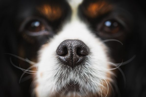
Your Dog’s Nose Is As Unique As A Fingerprint, And Its Distinct Pattern Actually Forms During Embryonic Development

Did you know your dog’s nose is as unique as your fingerprint? Each dog’s nose actually has its own distinct pattern of ridges and creases. These structures help keep the nose moist and aid in the sense of smell and temperature regulation.
Now, a new study has exposed how these patterns form during embryonic development. Researchers from the University of Geneva and several other institutions across Europe found that the “nose prints,” officially known as “rhinoglyphics,” form via a mechanical process.
The finding is significant because it has introduced scientists to the concept of “mechanical positional information.” It’s about how developing tissues need to know where to form certain structures, and they get this positional information from mechanical forces, not chemical signals.
“Finding specific examples of beautiful patterns in living organisms is easy,” said Michel Milinkovitch, a co-author of the study and a professor in Geneva’s Department of Genetics and Evolution.
“All we have to do is look around us! Our latest study focuses on the noses of dogs, ferrets, and cows, which exhibit a singular network of polygonal structures.”
The structures help dogs retain moisture on their noses. The moisture traps scent molecules, which can then travel to special sensory organs. The structures also assist with temperature regulation.
The research team used advanced imaging techniques to examine nose development in dog, cow, and ferret embryos with the aim of gaining a better understanding of how such intricate nose patterns form.
They discovered that the process consists of three primary stages. First, a network of blood vessels forms beneath the skin. The vessels are like the foundation of a building.
Next, the base layer of the skin, also known as the epidermis, starts to fold, creating cup-like structures between the vessels.

AnnaFotyma – stock.adobe.com – illustrative purposes only, not the actual dog
Finally, the outer layer of skin develops creases that line up with the blood vessels underneath, producing the unique, polygonal bumps we see on the noses of adult animals.
The most interesting part is that these patterns arise naturally from the physical properties of the tissues as they grow instead of following a predetermined genetic blueprint. The blood vessels act as guides, making sure the patterns are arranged in all the right areas.
The researchers confirmed the mechanism after making detailed observations and computer simulations.
They generated virtual models of growing nose tissue and demonstrated that the patterns emerged in their proper places when the blood vessels were stiffer than the surrounding tissue, just like how they were in real animals. The patterns became irregular and imprecise when the stiffness was taken out of the simulation.
“Our numerical simulations show that the mechanical stress generated by excessive epidermal growth is concentrated at the positions of the underlying vessels, which form rigid support points,” said Paule Dagenais, the first author of the study.
“The epidermal layers are then pushed outwards, forming domes—akin to arches rising against stiff pillars.”
It is the first time that mechanical positional information has been able to explain structural formation during embryonic development.
The researchers believe that the new principle of biological development could be linked to the formation of other biological structures and contribute to tissue engineering and biomaterial design innovations in the future.
The study was published in Current Biology.
Sign up for Chip Chick’s newsletter and get stories like this delivered to your inbox.
More About:Animals


